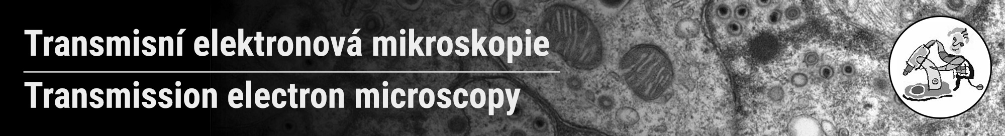
The Transmission Electron Microscopy laboratory (TEM) provides advanced expertise in cellular and material
electron microscopy: from sample preparation, surface imaging and high-resolution ultrastructure of samples, to analysis of the acquired data.
Available methods
- Standard chemical sample preparation
- Cryofixation of samples by high pressure deep freezing
- Automatic freeze substitution
- Preparation of semthini and ultrathin sections
- Preparation of cryosections by Tokuyasu method
- Preparation of samples for immunolabeling
- Antigen detection and localization by immunogold labeling
- Negative staining for surface morphology
We analyze
- Proteins and macromolecular complexes
- Viruses, exosomes, liposomes
- Bacteria and yeast cells
- Molds and plants
- Adherent and suspension cells
- Tissues
- Organoids and small organisms
- Nanoparticles
- Materials
Instrumentation
- Electron microscope JEOL JEM 2100
- High pressure deep freeze Leica EM PACT2
- Freeze substitution Leica EM AFS2
- Ultramicrotome with cryo chambre Leica EM UC7
Contact: Romana Hadravová, ☎ 136